Recently I attended a conference in London, England: Subluxation, Neurology and Health by Dr Dan Murphy and what I learned about neck posture, may shock you.
Why we need spinal curves
Early research by Bagnall in the Journal of Anatomy reported that the cervical lordosis (neck curve) begins to develop at just 9.5 weeks in utero and continues to develop when we first begin to hold our heads upright, at the approximate age of 3 months.
A newborn’s muscle strength is not sufficient to maintain the head in upright posture. Creating a better mechanical advantage in the neck can compensate for this lack of strength. The forward curving of the neck (lordosis) thus provides new muscle attachments and levers that offer a mechanical advantage, necessary to stabilize and move the head.
Reversed neck curves
When the ideal neck curve is lost or reversed (cervical kyphosis) – usually through trauma, there are cellular changes within the spinal cord that occur.
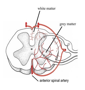
A 2005 study in the journal Spine, found that: “Progressive kyphosis of the cervical spine resulted in demyelination of nerve fibers,” and that these changes were associated with vascular changes in the spinal cord. Let me explain with a picture.

Above you see a cross section of the spinal cord which runs from the base of your brain, down through to the upper portion of your low back. If I were to take a slice out from the cord and look down over the slice, this is what I’d see.
Grey and white matter are both types of nerves. The former made up mostly of cell bodies and is grey in colour and the latter, made up of paler (more white) nerve fibers with their sheaths (covering) called myelin). It is the loss of this myelin that is associated with Multiple Sclerosis.
In the 2005 study, there was found to be significant correlation between the angle of the kyphosis (how much the neck curve was reversed) and the amount of spinal cord flattening. The greatest compression point was found to be at the apex of the bend (most commonly found on x-ray to be approximately C4-6).
The progression of demyelination
- trauma can cause a reverse neck curve
- a reversed neck curve compresses the spinal cord
- compression degrades the anterior spinal artery
- reduced blood flow leads to demyelination in the anterior (front) spinal cord, which controls motor function
- loss of motor control found in MS
There was severe demylination in the anterior funiculus … which controls posture and muscle tone. Shimizu et al.
The conclusion of the study was that a reversed neck curve (a kyphotic deformity) resulted in demyelination of the compressed white matter and affected the front of the spinal cord (supplied by the anterior spinal artery) which controls movement. As compression continued, so did the areas affected in the spinal cord.
Posture Analysis
One of the reasons that I insist on an x-ray analysis before treatment or exercise programs begin, is because of the potential for a reversed neck curve to influence systemic health.
Those of you with forward head posture, neck hump, chronic headaches or neck pain, should get a structural diagnosis (from x-ray) and if warranted, a tailored exercise program should be integrated, to help restore the normal (ideal) neck curve.
What is your experience – have you had any of the following signs or symptoms long-term?
- headaches
- neck pain
- visible fatty neck hump
- significant forward head leaning
- tingling/numb fingers or hands
- unexplained muscle weakness
- difficulty concentrating
- brain fog


